Pulmonary Valve Stenosis (PVS): Causes, Symptoms, Diagnosis, and Treatment
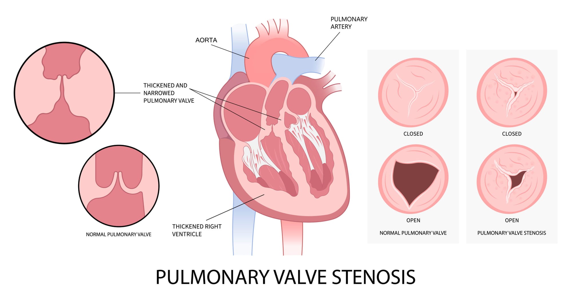
Pulmonary valve stenosis (PVS) is a type of heart valve disease that involves the narrowing of the pulmonary valve, which controls the flow of blood from the heart’s right ventricle into the pulmonary artery to carry blood to the lungs. During stenosis, the pulmonary valve’s flaps (also known as cusps or leaflets) are thickened, stiffened, or fused together. This narrowed opening of the valve slows or blocks the flow of blood into and through the pulmonary artery. Pulmonary stenosis can occur alone or in combination with other congenital heart defects. [1]
What are the 3 main types of PVS? [2]
VALVULAR
Valve itself is affected with varying degrees of fibrosis, thickening and commisured fusion causing restricted blood flow.
SUBVALVULAR
Obstruction present below the valve in the infundibular region of the right ventricle.
SUPRAVALVULAR
Also known as peripheral pulmonary stenosis, as the restriction can occur in the main pulmonary artery and/or its distal branches.
What are the causes of PVS?
PVS usually develops before birth when the pulmonary valve does not develop normally while growing inside the womb. The exact cause is still under research; however, a few conditions linked to the cause of congenital PVS may include:
Genetic syndromes such as Noonan syndrome, which is most commonly caused by PTPN11 mutations but can also be caused by KRAS, SOS1, and RAF1 mutations. [2]
Maternal rubella (German measles) is also a known cause of congenital valvular pulmonary stenosis, although it is not a genetic defect. [5]
Risk factors for non-congenital pulmonic stenosis include:
Carcinoid syndrome, which is a condition caused by carcinoid tumors in the digestive system that release chemicals into the bloodstream that may damage heart valves.
Rheumatic fever is a rare complication of strep throat.
Radiation to the chest, possibly from treatment for cancer close to the chest.
Rubella, a condition known as German measles that can increase the risk of pulmonary stenosis if experienced during pregnancy. [1]
What are the symptoms of PVS?
Mild pulmonary stenosis is rarely symptomatic at all. For moderate to severe PVS, the symptoms may include:
Chest pain
Shortness of breath
Dizziness or fainting
Fatigue
Bluish color to the skin or nail beds
Loss of appetite or struggle to gain weight in infants [3]
How is PVS diagnosed?
Echocardiography usually provides adequate visualization of the pulmonary valve and surrounding structures. Transesophageal echocardiography is used if suboptimal views or endocarditis are suspected.
Doppler studies using echocardiography provide flow gradients, which are used to grade the severity of the pulmonary stenosis. Guidelines from the European Association of Echocardiography, the American Society of Echocardiography, the AHA, the ACC, and the European Society of Cardiology (ESC) include the following severity categories:
Mild stenosis: The peak Doppler gradient across the pulmonary valve is <36 mm Hg, or the Doppler jet velocity is <3 m/s.
Moderate stenosis: The peak Doppler gradient across the valve is between 36 and 64 mm Hg, or the Doppler jet velocity is between 3 and 4 m/sec.
Severe stenosis: The peak Doppler gradient across the valve is >64 mm Hg, or the Doppler jet velocity is >4 m/s.
Electrocardiographic criteria that support right ventricular hypertrophy correlate with the severity of the pulmonary stenosis. Mild pulmonary stenosis may show right axis deviation, whereas severe cases may show prominent R waves in V1 and aVR, and P-wave enlargement. [2]
How is PVS treated?
For mild PVS, occasional health check-ups may be required to monitor symptoms. For moderate to severe PVS, surgical treatment may include:
Pulmonary valve replacement: If balloon valvuloplasty isn't an option, open-heart surgery or a catheter procedure may be done to replace the pulmonary valve.
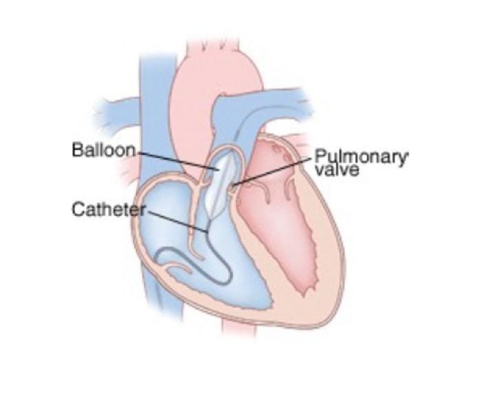
Balloon valvuloplasty: A balloon on the tip of a catheter is guided to the pulmonary valve via an artery. At the valvular junction, the balloon is inflated, enlarging the valve diameter. Narrowing can recur, and some people may need valve repair or replacement in the future.
People who have had pulmonary valve replacement need to take antibiotics before certain dental procedures or surgeries to prevent endocarditis. [4]
Share this post
Explore Related Articles for Deeper Insights
England Launches Emergency MenB Vaccination Programme for Students Ahead of 2026 Academic Year
Thousands of Young People to Receive Protection Against Meningococcal B Disease
The UK government h...