Tetralogy of Fallot: Causes, Symptoms, Diagnosis, and Treatment
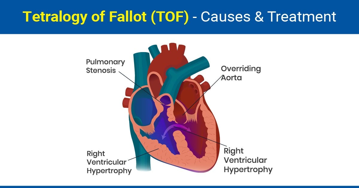
Tetralogy of Fallot (TOF) is a cardiac anomaly that refers to a combination of four related heart defects that commonly occur together. The four defects are:
Ventricular septal defect (VSD): A hole in the wall dividing the right and left ventricles.
Overriding aorta: the aortic valve is enlarged and appears to arise from both the left and right ventricles instead of the left ventricle as in normal hearts.
Pulmonary stenosis: Narrowing of the pulmonary valve and outflow tract or area below the valve that creates an obstruction (blockage) of blood flow from the right ventricle to the pulmonary artery
Right ventricular hypertrophy: thickening of the muscular walls of the right ventricle, which occurs because the right ventricle is pumping at high pressure. [1]
How common is TOF?
TOF is the most common cyanotic congenital heart disease, representing 5% to 7% of all congenital heart defects. [2]
About 1 in every 2,077 babies in the United States are born with tetralogy of Fallot. This means that about 1,768 babies are born with tetralogy of Fallot each year. [3]
What causes TOF in babies?
TOF occurs when a baby’s heart does not form correctly in the womb. Tetralogy of Fallot may be associated with chromosomal abnormalities, such as 22q11 deletion syndrome [2]. Children with certain genetic syndromes, such as Down`s syndrome or DiGeorge syndrome, may be at higher risk of developing TOF. Risk of getting TOF may be higher with:
Environmental factors, such as smoking or taking certain medications during pregnancy.
Family history
Having certain medical conditions during pregnancy, such as diabetes or rubella. [4]
What are the symptoms of TOF?
Tetralogy of Fallot symptoms depend on how much blood flow is blocked from leaving the heart to go to the lungs. Symptoms may include:
Blue or gray skin color
Shortness of breath and rapid breathing, especially during feeding or exercise.
Trouble gaining weight
Getting tired easily during play or exercise.
Irritability
Crying for long periods of time.
Fainting [5]
How is TOF diagnosed?
Some babies are diagnosed with tetralogy of Fallot before birth. The heart conditions may be seen and heard on prenatal ultrasounds, while the baby is still in the womb. Other tests that may help diagnose TOF after the baby`s birth may include:
Peripheral tissue oxygenation measurement by pulse oximetry.
Echocardiogram to check heart and heart valves and how well they are working.
Electrocardiogram to check irregular heartbeat or signals due to an enlarged heart.
A chest X-ray shows the shape and condition of the heart and lungs. A common sign of tetralogy of Fallot on an X-ray is a boot-shaped heart. That means the right lower chamber is too big.
Cardiac catheterization is occasionally required to evaluate the size and distribution of the pulmonary arteries. Catheterization can also demonstrate whether patients have pulmonary blood flow supplied by an abnormal blood vessel from the aorta (aortopulmonary collateral). [6]
How is TOF treated?
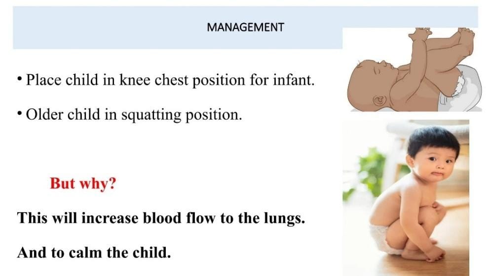
Once tetralogy of Fallot is diagnosed, the immediate management focuses on determining whether the child's oxygen levels are in a safe range. If the oxygen levels are too low, prostaglandin infusion is initiated to keep the ductus arteriosus open. This will provide additional pulmonary blood flow and increase the child's oxygen level. [2]
Occasionally palliative procedure to shunt more blood going to the lungs needs to be performed if the baby is too weak to undergo the open-heart surgical repair for TOF. [1]
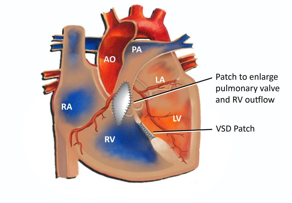
Children with TOF often need open heart surgery also known as complete repair within the first year of life. The surgeon patches the hole between the lower heart chambers and repairs or replaces the pulmonary valve. The surgeon may remove thickened muscle below the pulmonary valve or widen the smaller lung arteries.
After complete repair, the right lower chamber won't need to work as hard to pump blood. As a result, the right chamber wall should go back to its usual thickness. The oxygen level in the blood goes up. Symptoms typically get better. [6]
Share this post
Explore Related Articles for Deeper Insights
Young Colon Cancer Linked to Specific Fats in Ultra-Processed Foods: What New Research Reveals
New Study Suggests Certain Dietary Fats May Play a Role in Rising Colon Cancer Cases Among Young Adu...