Empyema: Causes, Symptoms, Diagnosis, and Treatment
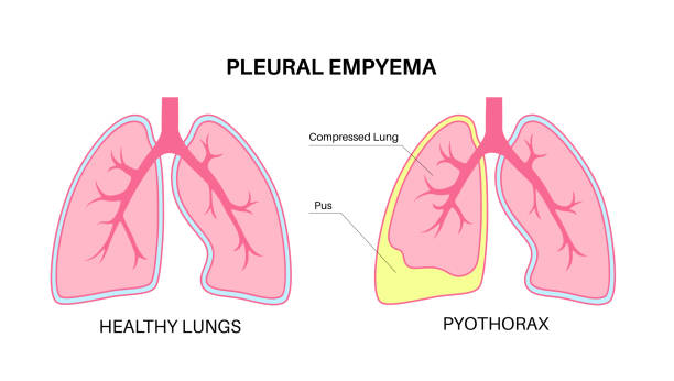
Thoracic empyema is an infectious condition in which pus develops in pleural space. This condition was first recognized by Hippocrates and is historically associated with high mortality rates.
How common is empyema?
Pleural infections, including empyema and complicated parapneumonic effusion, pose a serious health risk, impacting approximately 80,000 patients annually in the United States (US) and the United Kingdom (UK). These infections carry a 30-day mortality rate reaching up to 10.5% and exceeding to 19% at 1 year. Furthermore, more than half of patients likely develop parapneumonic effusions, resulting in increased morbidity across all patient groups, from those with simple effusions to those with advanced empyema. Beyond mortality, these infections impose significant morbidity, as patients often experience prolonged hospital stays—averaging 19 days, according to recent reviews—and frequently present with multiple comorbid conditions.
What is the estimated economic burden of empyema?
The estimated average annual expenditure associated with thoracic empyema is $500 million. A study by Mummadi et al reveals a rise in empyema hospitalizations in the US from 2007 to 2016, with in-hospital mortality ranging from 5.2% to 6.2% and an average cost per hospitalization of $38,591. These factors underscore the need for refined diagnostic and treatment strategies to improve patient outcomes and alleviate the economic impact of thoracic empyema. [1]
What are the causes of empyema?
The most common causes of empyema include [2]:
Bacterial pneumonia
Tuberculosis
Chest surgery
Lung abscess
Trauma or injury to the chest
The following are the risk factors to increase your likelihood of getting empyema:
Age older than 70.
Any recent surgeries of the thoracic region.
Having Diabetes.
Having chronic obstructive pulmonary disease (COPD).
Having bronchiectasis.
Having a blood clot.
Using intravenous injections for drugs on a regular basis.
What are the symptoms of empyema?
Dry cough
Excessive sweating, especially night sweats
Fever and chills
General discomfort, uneasiness, or ill feeling (malaise)
Shortness of breath
Weight loss (unintentional) [3]
Pleurisy, chest pain that worsens with deep breathing
What are the stages of empyema?
The American Thoracic Society first described the evolution of empyema as a continuous process subdivided into 3 distinct but continuous stages or phases:
Exudative stage: Characterized by rapid fluid accumulation in the pleural space with Gram negative stained culture and glucose levels exceeding 60 mg/dL. Proinflammatory mediators like tumor necrosis factor-alpha (TNF-α), interleukin 6 (IL-6), and IL-8 are believed to be vital in driving the inflammatory response at this stage.
Fibrinopurulent and loculated stage: This phase is characterized by bacterial invasion into the pleural space, often as a result of inadequate or delayed antibiotic treatment. In this stage, the pleural fluid will generally have a glucose level below 60 mg/mL with a pH below 7.20 and a pleural fluid LDH greater than 3 times the upper limit of normal for serum. Increased plasminogen-activator inhibitors and TNF-α lead to fibrin deposition, which forms septations and loculations. Within these loculations, walled-off bacteria increase neutrophil phagocytic activity, a corresponding increase in LDH, and increased production of lactic acid and glucose consumption; this explains the changes observed in pleural fluid analysis.
Chronic organizational stage: This stage is marked by the growth of fibroblasts into the visceral and parietal pleurae and is accompanied by the deposition of a collagen-rich fibrin matrix within the pleural space, resulting in pleural thickening. Development of an inelastic visceral pleural peel can cause lung entrapment. [1]
How is empyema diagnosed?
Pleural ultrasonography: Can identify small pleural effusions and may be better than Chest X-rays at estimating the volume of the effusion, while not being hindered by adjacent consolidations or intrathoracic structures.
Chest computed tomography (CT): Contrast-enhanced CT with tissue phase is a valuable imaging tool in the assessment of pleural-space infections. In addition, it allows a detailed examination of parenchymal abnormalities and may reveal a cause for the pleural space infection, such as bronchogenic carcinoma, endobronchial foreign body, or esophageal rupture.
Pleural fluid analysis and biomarkers: In general, purulence of the pleural fluid or a positive Gram's stain or culture from the pleural fluid establishes the diagnosis of empyema and should prompt tube thoracostomy drainage. A pleural fluid pH of <7.2 OR pleural fluid glucose value <40 mg/dL OR pleural fluid LDH value >1000 IU/L should prompt chest tube drainage.
Pleural fluid culture: Culture specimens should be obtained in all cases of acute bacterial empyema. It is recommended that culture specimens are acquired during aspiration or drainage procedures. [4]
Blood tests indicate signs of infection, such as high white blood cell count, C-reactive protein, and can help identify causative pathogens and bacteraemia.
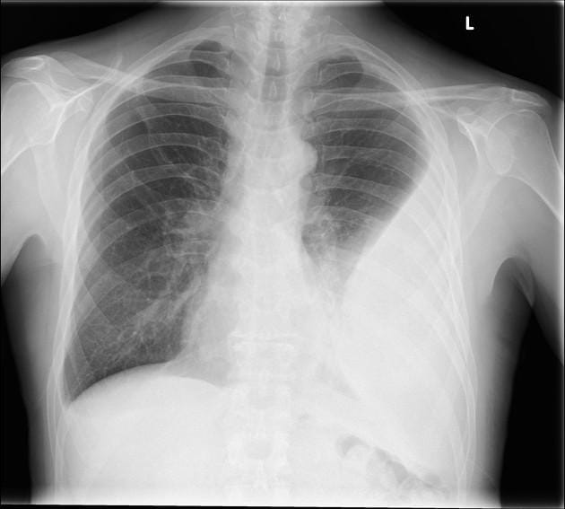
Chest X-ray: Conventional CXR is often the most available imaging modality leading to the identification of a pleural space infection. A minimum of 175 mL of pleural fluid is necessary to result in blunting of the costophrenic angle on a posteroanterior film, although smaller effusions may be identified on a lateral view.
What is the treatment for empyema?
Treatment involves removing pus through thoracentesis in the early stages of empyema. If drainage isn’t enough, a provider may try to break up the pus through fibrinolytic therapy.
In the later stages of empyema, a provider may need to perform a more invasive procedure, such as surgically removing fibrous tissue (decortication), a thoracotomy, or a video-assisted thoracic surgery (VATS).
Antibiotics to treat mild cases of empyema include, but are not limited to, the following [2]:
Amoxicillin-clavulanate
Piperacillin-tazobactam
Imipenem
Meropenem
Share this post
Explore Related Articles for Deeper Insights
New Cancer Injection Shows Promise in Eliminating Tumors After Other Treatments Fail
Groundbreaking Cancer Therapy Delivers Remarkable Results in Global Clinical Trial
A new cancer tre...