Pulmonary emphysema, a progressive form of chronic obstructive pulmonary disease (COPD), is characterized by persistent respiratory symptoms and airflow limitation resulting from airway or alveolar damage.
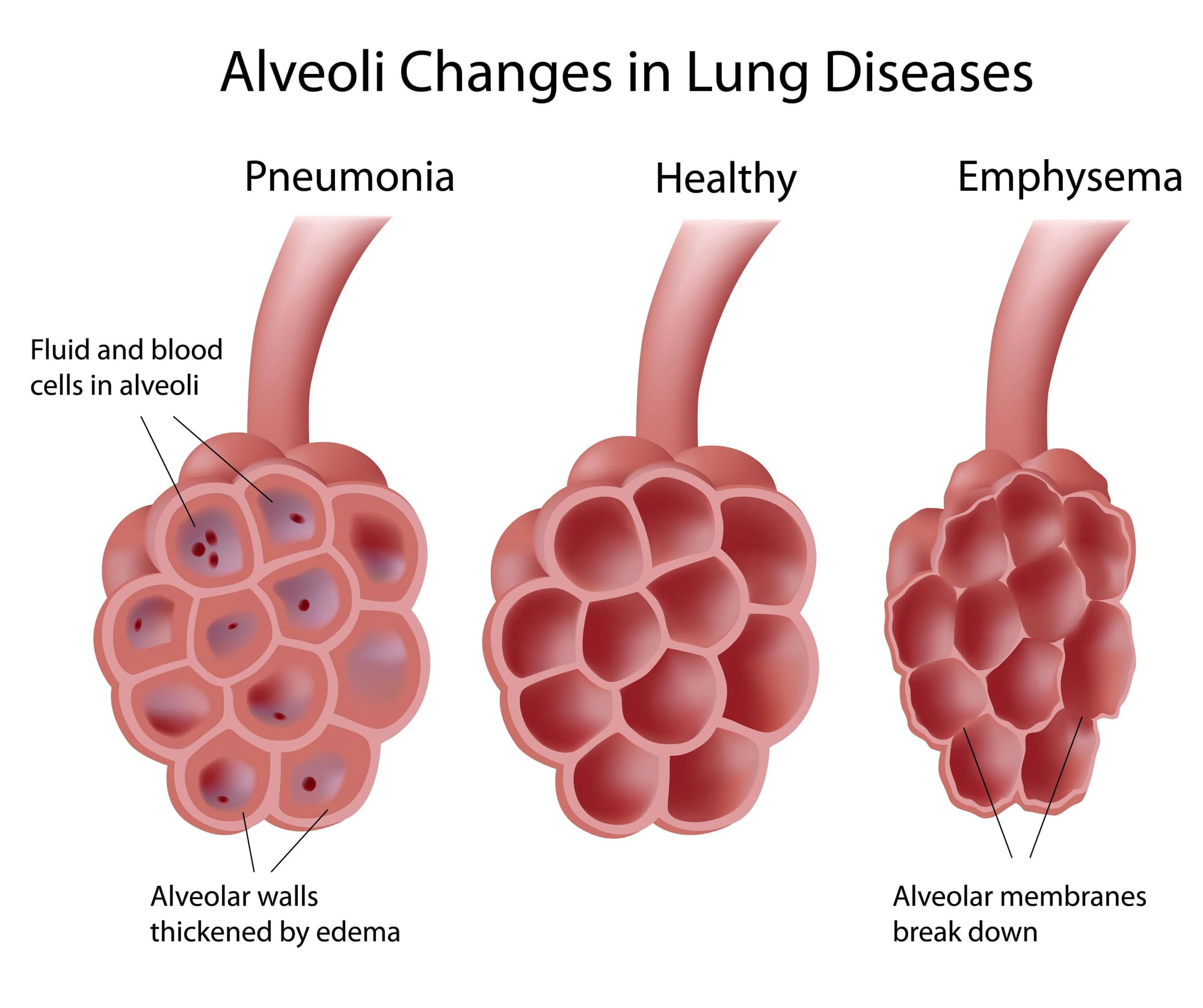
Alveoli are small, thin-walled, fragile air sacs arranged in clusters at the end of the bronchial tubes (airways) deep inside your lungs. There are about 300 million alveoli in adult lungs. The alveoli are where the lungs and the blood exchange oxygen and carbon dioxide during the process of breathing in and breathing out. Oxygen breathed in from the air passes through the alveoli and into the blood and travels to the tissues throughout the body. Carbon dioxide travels in the blood from the body's tissues and passes through the alveoli to be breathed out.
What are the types of Emphysema?
The clinical manifestations of emphysema arise from damage to the airways distal to the terminal bronchiole, including the respiratory bronchioles, alveolar sacs, alveolar ducts, and alveoli, collectively known as the acinus. In emphysema, there is abnormal permanent dilatation of the airspaces and destruction of their walls due to the proteinase activity. This dilatation decreases the alveolar and capillary surface area, impairing gas exchange. The specific part of the acinus affected determines the subtype of emphysema. Emphysema can be pathologically subdivided into the following types:
Centrilobular (proximal acinar) emphysema is the most common type, typically associated with smoking. It can also be observed in individuals with coal workers' pneumoconiosis.
Pan acinar emphysema is most commonly associated with alpha-1 antitrypsin deficiency.
Para-septal (distal acinar) emphysema may occur alone or in combination with the other 2 types. When it occurs independently, it is often associated with spontaneous pneumothorax in young adults. [1]
What are the symptoms of Emphysema?
Many people don’t notice emphysema symptoms until the disease has destroyed 50% or more of their lung tissue. Until then, the first signs include gradual shortness of breath and fatigue. Other symptoms may include:
Long-term coughing (smoker`s cough)
Wheezing
Shortness of breath, especially during light exercise like climbing steps.
Constant feeling of not being able to get enough air.
Tightness in your chest
Increased mucus production
Abnormal mucus color (yellow or green)
Ongoing fatigue
Heart problems
Trouble sleeping
Anxiety and depression
Weight loss
Having emphysema can increase the risk of getting pneumonia, bronchitis and other lung infections.
What are the causes of emphysema?
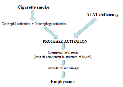
Cigarette smoking is the main cause of emphysema. Smoking causes inflammation, damaged cilia, swollen airways, mucus production and difficulty clearing your airways. Other causes of emphysema include:
Marijuana
Vaping and e-cigarettes
Cigar smoke or being around second-hand smoke
Toxins in the air
Dust
Chemical fumes [2]
How is emphysema diagnosed?
Following tests not only help diagnose emphysema, but also track disease progression:
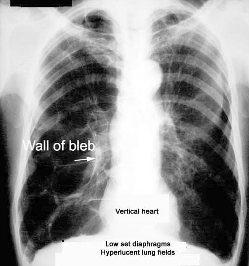
X-rays may show some changes in the lungs caused by emphysema; however, a CT scan gives much greater detail of changes in your lungs than a chest X-ray does.
Spirometry is the most common lung function test to measure how much air your lungs can hold and how fast you can blow the air out of your lungs. Spirometry reveals air flow limitations.
Other tests include measurement of lung volumes and diffusing capacity, the six-minute walk test, and pulse oximetry.
Arterial blood gas analysis does not help diagnose emphysema, but it reveals how well your lungs are bringing oxygen into your blood and removing carbon dioxide.
Blood tests can also be done to check the genetic deficiency of alpha-1-antitrypsin, which contributes to the pathophysiology of emphysema.
How is emphysema managed conservatively?
The most important step in any treatment plan for emphysema is to quit all forms of smoking. Pharmacotherapy treatment for emphysema includes:
Bronchodilators: These usually come in the form of inhalers. Bronchodilators relax the muscles around your airways. This can help relieve coughing and make breathing easier. Depending on the severity of your emphysema, you may need a short-acting bronchodilator before activities, a long-acting bronchodilator that you use every day, or both.
Inhaled steroids: These help reduce airway inflammation and keep exacerbations from happening. Side effects may include bruising, mouth infections, and hoarseness. These medicines are useful if you often have exacerbations of emphysema.
Combination inhalers: include one or more bronchodilators and steroids.
Antibiotics: are also prescribed to treat bacterial infection with co-existing bronchitis or pneumonia.
Oral steroids: A short course can be prescribed during exacerbation to prevent symptoms from getting worse.
Pulmonary rehabilitation: Involves working with different professionals to improve awareness and education about managing the symptoms, exercise training, draining of secretions, nutrition advice, and counseling.
Supplemental oxygen: In severe emphysema, blood oxygen levels can be low. Lightweight, portable units can help you breathing during physical activity and help you sleep better.
Alpha-1-antitrypsin deficiency: Some people can be treated by also replacing the missing AAT protein. This may stop more damage to the lungs.
What are the surgical options for emphysema?
Lung volume reduction surgery: Small wedges of damaged lung tissue from the upper lungs is removed by the surgeon. This creates extra space in the chest so that the remaining healthier lung tissue can expand and the muscle that helps in breathing can work better
Endobronchial valve surgery: A minimally invasive procedure in which a tiny one-way endobronchial valve is placed in the lung. Air can leave the damaged part of the lung through the valve, but no new air gets in. This allows the most damaged lung lobe to shrink so that the healthier part of the lung has more space to expand and function.
Bullectomy: Bullae are large air-filled spaces in the lungs that are formed when the inner walls of the alveoli are destroyed. Large bullae can reduce the lung surface area and cause breathing problems. In a bullectomy, the surgeon removes the bullae from the lungs to allow more air flow
Lung transplant: This is the last option indicated when all other treatment options are unable to achieve the desired improvement in symptoms. [3]
Share this post
Explore Related Articles for Deeper Insights
Turmeric Benefits, Uses, Side Effects, and Safety: A Complete Guide
Why Turmeric Remains One of the Most Popular Natural Health Supplements
Turmeric has been used for ...